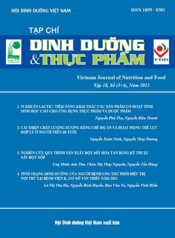
CASE STUDY: IMPROVE BONE QUALITY WITH A PROPER DIET AND EXERCIS, IMPLEMENTED IN 4 YEARS FOR ONE PERSON > 60 Y OLD
Main Article Content
Abstract
Aims: To evaluate the effects of proper dietary and exercise practice on bone mineral density mass (BMD), bone quality index (BQI), and broadband ultrasound attenuation (BUA) in a person aged 60 years.
Methods: The daily diet provides 2000Kcal, 1200mg Ca, 1.7mg protein/kg of body weight, 3800IU vitamin D, etc. Exercise 3 times/week x 60 minutes/time with moderate and heavy intensity; 45minutes daily with moderate and light intensity, and 8 hours working at office. Bone quality measured by QUS instrument every 6 months.
Results: At the beginning of the intervention, the T-score of heel bone was -2.4 (64.5%, high risks of osteoporosis), and increased to 0.8 (113% of good level) at the end of the intervention. Other indicators such as BQI and BUA also improved from 69.1pt & 39.5dB/MHz at the beginning to 121.1 pt (+95.6%) and 108.1dB/MHz (+173.7%) at the 48th month, respectively.
Conclusion: A proper diet and exercise improved bone quality recovery in the elders.
Keywords
Older person, calci, exercise, osteoporosis, QUS, BQI, BUA
Article Details
References
2. WHO. Diagnosis and assessement. In: Prevention and management of osteoporosis. Geneva 2003. Pp 53-81.
3. Nguyễn Thị Ngọc Lan, Hoàng Hoa Sơn. Khảo sát yếu tố nguy cơ loãng xương ở phụ nữ Việt Nam từ 50 tuổi trở lên và nam giới từ 60 tuổi trở lên. Nghiên cứu Y học. 2015;5:91-98.
4. WHO. WHO guidelines on physical activity and sedentary behaviour: at a glance. Geneva 2020. Pp 4-6.
5. Dionyssiotis Y, Paspati I , Trovas G, et al. Association of physical exercise and calcium intake with bone mass measured by quantitative ultrasound. BMC Women’s Health. 2010, 10:12. doi:10.1186/1472-6874-10-12.
6. Bộ Y tế/Viện Dinh dưỡng. Nhu cầu dinh dưỡng khuyến nghị cho người Viêt Nam. NXB Y Học Hà Nội 2016.
7. Li C, Sun J, Yu L. Diagnostis value of calcaneal quantitative ultrsound in the evaluation of osteoporosis in milddle-aged and elderly patients. Medicine. 2022;101:1-6.
8. Zhu ZQ, Liu W, Xu CL, et al. Ultrasound bone densitometry of the calcaneus in healthy Chinese children and adolescents. Osteoporos Int. 2007;18:533–541.
9. Prins SH, et al. The role of quantitative ultrasound in the assessement of bone: a review. Clinical Physiology. 18;1:3-17.
10. Langton CM, Njeh CF, Hodgskinson R. Et al. (1996) Prediction of mechanical properties of the human calcaneus by broadband ultrasonic attenuation. Bone. 1996;18: 495±503.
11. Srichan W et al. Bone status measured by quantitative ultrasound: a comparison with DEXA in Thai children. Eur J Clin Nutr. 2015, 1–4. doi:10.1038/ejcn.2015.180
12. Albanese CV, De Terlizzi F, Passariello R. Quantitative ultrasound of the phalanges and DXA of the lumbar spine and proximal femur in evaluating the risk of osteoporotic vertebral fracture in postmenopausal women. Radiol Med. 2011;116:92–101.
13. Gluer CC. Quantitative ultrasound techniques for the assessment of osteoporosis: Expert agreement on current status. J Bone Miner Res. 1997;12:1280-1288.
14. Baran DT. Quantitative ultrasound: a technique to target women with low bone mass for preventive therapy. Am J Med. 1995; 98(2A):48S-51S.
15. Bennell KL, Hart P, Nattrass C, Wark JD. Acute and subacute changes in the ultrasound measurements of the calcaneus following intense exercise. Calcif Tissue Int. 1998;63:505-509.
16. Pinheiro MB , Oliveira J, Bauman A et al. Evidence on physical activity and osteoporosis prevention for people aged 65+ years: a systematic review to inform the WHO guidelines on physical activity and sedentary behaviour. Inter J Behav Nutr Phys Act. 2020;17:150-159.
17. Prince R, Devine A, Dick I, et al: The effects of calcium supplementation (milk powder or tablets) and exercise on bone density in postmenopausal women. J Bone Miner Res. 1995;10:1068-1075.
18. Nelson ME, Fisher EC, Dilmanian FA, et al. A 1-y walking program and increased dietary calcium in postmenopausal women: Effects on bone. Am J Clin Nutr. 1991;53:1304-1311.
19. Nelson ME, et al.. Effects of high-intensity strength training on multiple risk factors for osteoporotic fractures. JAMA. 1994;272:1909-1914.
20. Jones PRM et al. Influence of brisk walking on the broadband ultrasonic attenuation of the calcaneus in previously sedentary women aged 30-61 years. Calcif Tissue Int. 1991;49:112-115.
21. Hoshino H, Kushida K, Yamazaki K, et al. Effect of physical activity as a caddie on ultrasound measurements of the os calcis: a cross-sectionalcomparison. J Bone Miner Res. 1996;11:412-418.
22. Heinonen A, Kannus P, Sievanen H, et al. Randomised controlled trial of effect of high-impact exercise on selected risk factors for osteoporotic fractures. Lancet. 1996;348:1343-1347.


